Description:
MRI method that allows faster data acquisition with lower noise and higher accuracy, with application in stroke, brain tumours and dementia
Background
MRI technology has revolutionised healthcare and is currently the leading method for clinical and research studies of the brain and other biological systems. Worldwide, there are more than 50,000 MRI scanners machines installed performing around 95 million scans per year. With the growing awareness about the benefit of early diagnosis, the rising demand for non-invasive procedures and the increase of the geriatric population there is a need for the development of well validated and robust quantitative imaging biomarkers.
Some MRI neuroimaging methods are Diffusion Weighted Imaging (DWI), Diffusion Tensor Imaging (DTI) and Diffusion Kurtosis Imaging (DKI). Although their many benefits, these methods have limitations regarding acquisition times and accuracy.
For new MRI techniques to be clinically useful there is a need for rapid image acquisition to ensure patient comfort, maximise image quality (by minimising time for patient movement) and to minimise the economic cost. This means there is a requirement to obtain images with excellent signal to noise tissue contrast at short acquisition times.
Technology Overview
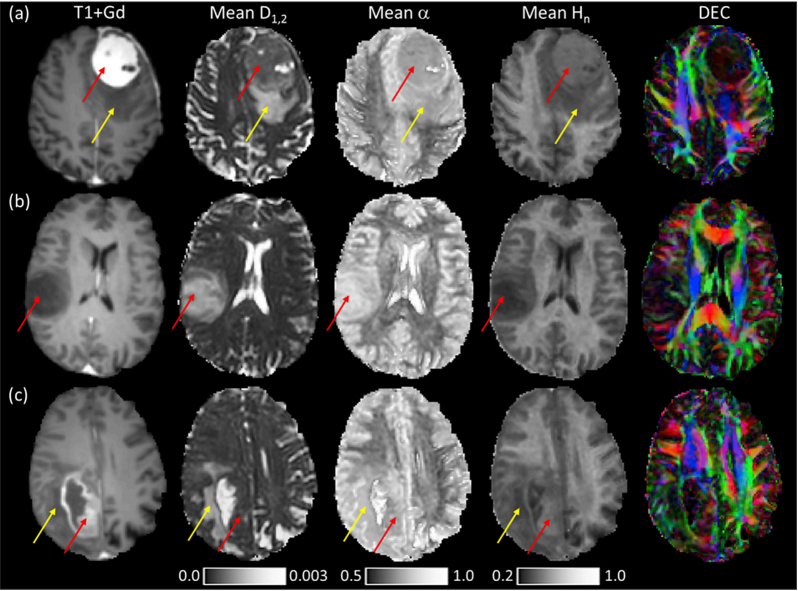
Researchers at St. Georges University of London have developed a novel quantitative MRI method, called Quasi-Diffusion Imaging (QDI), that provides high quality images with accurate and precise parameter estimation from an optimised protocol. It utilises an extremely fast imaging protocol with high contrast and signal to noise ratios that provides high tissue contrast images. With applications in stroke, brain tumours and dementia, the method can quickly and accurately detect microstructural damage in disease.
QDI overcomes other neuroimaging methods limitations and provides DWI, DTI and DKI maps as well as novel diffusion parameter map. Providing additional diagnostic or prognostic information beyond that of conventional DWI and DTI, this new quantitative MRI technique can be acquired on a standard clinical MR system in a clinically acceptable time and can guide future therapeutic and intervention strategies.
Benefits
QDI is a fast image protocol that can be added to routine conventional MRI acquisitions allowing simple application in clinical trials and translation to the clinical arena.
It generates images with superior tissue contrast to conventional diffusion imaging, by fitting acquired data more stably in the presence of noise and with clinically acceptable acquisition times of between 84 and 228 s.
With applications in stroke, brain tumours and dementia, this novel method provides a rapid technique for identifying acute stroke lesions in the clinic including valuable information regarding patient recovery. And it also provides clinically meaningful images in cerebral small vessel disease and brain tumour case studies.
Applications
The QDI method has a particular advantage in aiding clinical staff in quickly and accurately detecting microstructural damage in disease, and has clear applications in stroke, brain tumours and dementia. Although it can also be used in other biological systems.
This method can also be used in routine clinical work and implemented on standard clinical MRI systems, as it overcomes the acquisition time limitations of other methods allowing clinically feasible image acquisition.
Opportunity
St. Georges University is looking for MRI scanner manufactures or clinical MRI analysis software companies interested in licensing this technology.
Patents
The Quasi-Diffusion Imaging method patent is pending in EU, US and JP.