Description:
This invention provides a novel method of analysing MRI data of a tumour in order to determine the tumour type.
Background
Glial tumours are the most common primary brain tumour and exhibit a heterogeneous infiltrative growth pattern. Although biopsies are still key to assessing tumour type and grade, they do not provide a full 3D picture of the tumour characteristics. Conventional MRI provides 3D images with an indication of the tumour core and overall extent, but is not optimal for determining the tumour grade nor distinguishing the true extent of tumour infiltration into surrounding brain. Thus conventional MRI is limited in providing accurate information for individualized radiotherapy planning, that can lead to both under and over-treatment of the lesion in terms of radiotherapy dose-planning. These shortcomings are apparent in the eventual recurrence of gliomas post-treatment and in radiation effects in normal brain.
Therefore, there is a great need for a non-invasive method that can aid in grading glial brain tumours and distinguish between healthy and cancerous tissue.
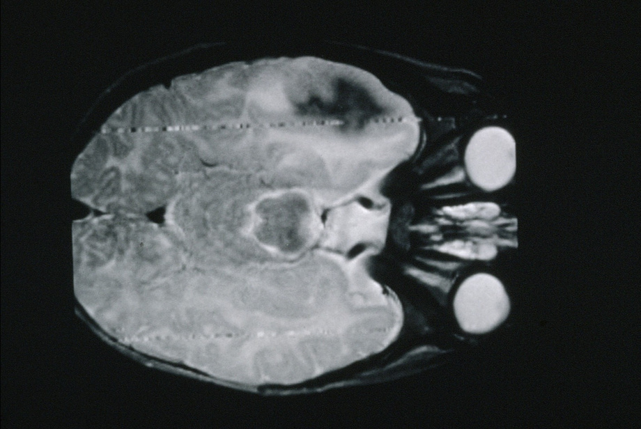
Technology Overview
A method of tissue-type mapping of glial tumours using multimodal magnetic resonance (mMRI) has been developed to provide 3D maps of the low or high grade tumour core and necrosis, the extent of tumour infiltration, and the presence of oedema. The statistical combination of data from multiple MRI types enables high specificity for detecting these tissue types that is not possible with the individual MRI methods alone. In the current implementation magnetic resonance spectroscopic imaging (MRSI) has been used to define regions of specific tissue types from which MR imaging characteristics are obtained. Information from quantitative MR diffusion and relaxation time parameters are then combined in 4D space to provide statistical priors for tissue-type probability density distributions (PDDs). A Bayesian classification scheme can then be used to generate whole brain tissue probability maps from mMRI datasets of individual patients.
Benefits
This analysis method combines mMRI to provide tissue classification and thus determine the grade, spatial extent and heterogeneity of glial brain tumours with proof-of-principle in a cohort of 25 low grade and high grade glioma patients.
Using an expandable Bayesian analysis of mMRI data this novel technique is not limited by the underlying assumptions of Gaussian statistics and does not require tissue spatial priors – a particular advantage in tissue segmentation of tumours that are heterogeneous in location and presentation.
Tumour tissue-type mapping with multimodal MRI may aid management of patients with brain tumours by delineating viable tumour from oedema and normal brain to aid targeted biopsy, surgical planning and radiotherapy dose-planning, as well as prognosis for patient survival.
Applications
This is a mathematical method for using multimodal MRI to infer the likely tissue-type of brain tumours, but which can be readily expanded to include other imaging modalities and tissue-types to further improve and extend its capabilities.
Opportunity
Licensing and partnering.
Patents
- WO2014174317A2
Seeking
- Licensing
IP Status
- Patented